
There is a bursa (fluid filled sac) that lies between the popliteal tendon and the posterior surface of the knee joint – it is called the popliteus bursa. The popliteus is located behind the knee joint, forming the base of the popliteal fossa. The remaining three muscles (tibialis posterior, flexor hallucis longus and flexor digitorum longus) act on the ankle and foot. One muscle, the popliteus, acts only on the knee joint. There are four muscles in the deep compartment of the posterior leg. Actions: Contributes to plantarflexion at the ankle joint and flexion at the knee joint.It blends with the calcaneal tendon and inserts onto the calcaneus. The fibres condense into a tendon which travels down the leg, between the gastrocnemius and soleus muscles. Attachments: Originates from the lateral supracondylar line of the femur.The plantaris is a small muscle which is absent in approximately 10% of the population. Actions: Plantarflexion of the foot at the ankle joint.The muscle converges with the fibres of the gastrocnemius to form the calcaneal tendon, which inserts onto the calcaneus. Attachments: Originates from the soleal line of the tibia and proximal fibula.

It gets its name from its resemblance to a sole – a flat fish. The soleus is a flat muscle located underneath the gastrocnemius.
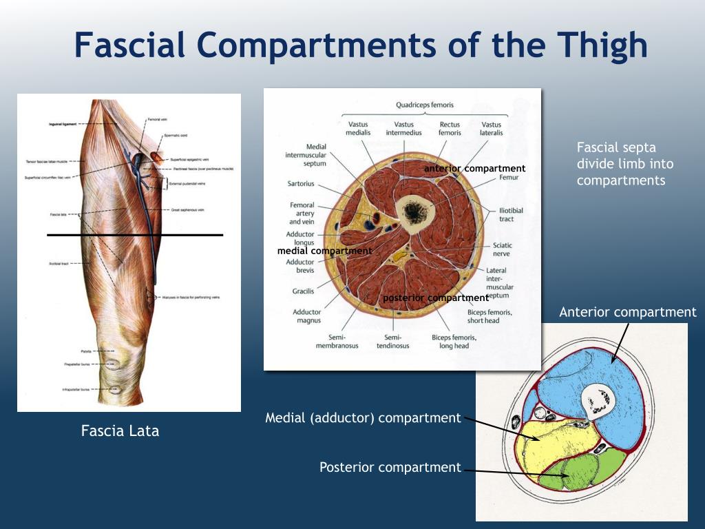
FASCIAL COMPARTMENTS OF LEG SKIN
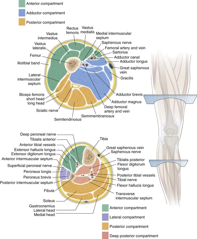
Subcutaneous calcaneal bursa – lies between the skin and the calcaneal tendon.To minimise friction during movement, there are two bursae (fluid filled sacs) associated with the calcaneal tendon: They all insert onto the calcaneus (heel bone) of the foot, via the calcaneal tendon. The anterior crural septum divides the muscles in front of and lateral to the two bones into an anterior compartment, which contains four muscles including tibialis anterior, and a more laterally placed peroneal compartment, which contains two of the three peroneal muscles.There are three muscles within the superficial compartment of the posterior leg. These two septa are attached to the fibula here, and here. This is the posterior crural septum, lying just in front of the soleus muscle. To see them, we’ll remove the investing deep fascia down to here, exposing several muscles that we haven’t met yet. The other two septa have cumbersome names: they’re the anterior and the posterior crural intermuscular septa. At the ankle, the transverse septum is continuous with the flexor retinaculum. The transverse septum is thin up here, but toward the ankle it becomes thicker. To see the transverse septum better, we’ll remove the rest of soleus.

Three muscles that we haven’t seen yet lie between the transverse septum and the bones. It runs from here on the tibia, to here on the fibula. In front of soleus, this transverse intermuscular septum crosses the back of the leg. Here’s soleus, divided, here’s the investing deep fascia, divided at a lower level. We’ll remove gastrocnemius and soleus, down to here. Together with the interosseous membrane, they divide the muscles of the leg into four compartments, two on the front of the leg, and two on the back. Now we’ll look at the fibrous septa, the singular of which is septum. Distally the investing deep fascia is continuous with the superficial part of the flexor retinaculum, with the peroneal retinaculum, and with the two parts of the extensor retinaculum. The investing deep fascia wraps around the back of the calcaneal tendon, like a sling. It’s attached to the fibula not directly, but indirectly by two fibrous septa here, and here, that we’ll see in a minute. The investing deep fascia is attached to the tibia here, and here. This outer layer is the investing deep fasica. Before we move on to see the muscles that produce inversion and eversion, we need to digress for two minutes, to look at the layer of deep fascia that surrounds all the muscles of the leg, and the three fibrous partitions, or septa that divide the leg muscles into somewhat distinct compartments.


 0 kommentar(er)
